Monday, October 22, 2018

Determination of truth from deception using functional MRI and cognitive engrams

Determination of Truth from Deception Using Functional MRI and Cognitive Engrams

D Marks, M Adineh, S Gupta

The Internet Journal of Radiology. 2005 Volume 5 Number 1.

http://ispub.com/IJRA/5/1/9241

Abstract

INTRODUCTION Investigators have attempted for centuries to determine the truthfulness and accuracy of statements (Ford 2006). Methodologies include standard interrogation, with and without use of medication or coercion, polygraph, and brain fingerprinting using brain evoked response potentials (ERP). None of these technologies yields optimal results, for various reasons, and are not entirely suitable for measuring deception.

An association between the ERP and lying on a standard test such as the Guilty Knowledge Test (GKT) suggested that deception may be associated with changes in other measures of brain activity such as regional blood flow (RBF) (Langleben 2002; Mochizuki 2001). Changes in RBF, as Blood Oxygen Level Dependent (BOLD) can be monitored using functional magnetic resonance imaging (fMRI), and have been shown to be measurements of regional brain activity (Ogawa 1990). These areas of brain activation on fMRI have been correlated with active memory encoding (Wegner 1999).
A number of researchers (Slotnick et al, Langlaben et al, Lee et al, Kozel et al) have sought to apply fMRI to discern between truth and deception. In general, test subjects were presented with artificial situations during which they were given the opportunity to tell the truth or give a false response to questions, while undergoing fMRI. The design of presentation of questions were different between research groups. These separate research groups were able to detect truthful or deceptive responses, based upon analysis of activation maps showing where in the brain there was increased neural activity. The areas of activation corresponding to truth or deception differed among groups of researchers, making a universal framework for analysis of interrogation with fMRI difficult to develop.

The purpose of this study was to validate prior work of other investigators, and to develop a paradigm to address interinvestigator variability that needs to be resolved before fMRI can be applied to interrogation in practice. Our methods employ activation area analysis, both for truth and deception, and also use activation map patterns for object recognition. These combination analytical methods should yield a more robust schema for use of fMRI as an interrogation technique. One of the central means of analysis we employ is the concept of the cognitive engram, which we define as the multi-dimensional representation of activation areas in the brain, which we contend represent the actual specific thought process. We have grouped truthful and deceptive responses, and also the recognition of specific faces and objects, into cognitive Engrams. Certainly when an individual thinks of a truthful or a deceptive response, or recognizes a face or an object, that concept transforms into the more generalized concept of truthfulness or deception, or recognition of a specific person. This overall recognition is not simply fragmented into hundreds of isolated activation points, but should be treated as a whole, as a cognitive engram, consisting of a number of widely distributed activation areas representing a single concept.

METHODOLOGY We operated under an IRB-approved protocol, and all volunteers signed an informed consent form. Healthy adult volunteers without exclusions to MRI scanning were explained the study purpose and design, risks and benefits, given a chance to ask questions and then agreed to participate. Continual neuroimaging (fMRI) was performed during viewing of the test stimuli in order to capture structural and functional data. The data was analyzed for the presence of neuroimaging activation that has been shown to correspond to cognition and visual recognition.

Cardiomyopathy due to ingestion of Adderall


Cardiomyopathy Due to Ingestion of Adderall


American Journal of Therapeutics 15, 287–289 (2008)


Donald H. Marks, MD, PhD*

A patient is described who developed cardiomyopathy after receiving a therapeutic course of dextroamphetamine/amphetamine. The patient’s cardiac function deteriorated to the point of heart failure, necessitating a heart transplantation. Cardiomyopathy associated with amphetamine use is a serious and potentially lethal condition. With early diagnosis, identification of the cause, and treatment, cardiomyopathy may be reversible. The dangers of therapeutic use of amphetamines are discussed, as well as problems and assumptions associated with U.S. Food and Drug Administration monitoring and removal from the market of harmful substances.

Keywords: Adderall, Ritalin, dextroamphetamine, amphetamine, cardiomyopathy
INTRODUCTION
Amphetamines are noncatecholamine sympathomi- metic amines with CNS stimulant activity.1 As prescrip- tion drugs, they are prescribed widely for attention deficit and hyperactivity disorders.2 Of the number of adverse effects that are associated with amphetamines, one that can occur, sometimes with devastating results, is cardiovascular toxicity.3 I describe a case of cardio- myopathy leading to a heart transplantation which was causally related to the prescription drug Adderall.
METHODS
The basis of this case report was a thorough review of the medical records, a medical history and physical examination after the heart transplantation had oc- curred, and a search of the current medical literature using Medline. A determination of causal relatedness between the consumption of Adderall (Shire Pharma- ceuticals, Wayne, PA) and the development of cardio- myopathy was made by means of universally accepted algorithms,4 as described.
 Case report
The patient was a 34-year-old Caucasian man with a medical history of asthma, obesity, irritable bowel syn- drome, and genital herpes. He was given a diagnosis of attention deficit disorder and was prescribed and ingested Adderall (dextroamphetamine/amphetamine) for approximately a 2-year period.
Adderall was started initially at 20 mg daily. Two months later the dosage of Adderall was increased to 40 mg a day, and Prozac, 20 mg, was added as a mood stabilizer. Seven months later the Adderall dosage was again increased to 40 mg in the morning and 20 mg at night, as well as an increase in Prozac to 40 mg each day. The next month, the dose of Adderall was again increased, to 80 mg—three 20-mg tablets in the morning and one tablet in the evening—because the patient was still having problems with completing tasks. On the higher dose of Adderall, the patient experienced increased hyperactivity and irritability, and in response to these symptoms his neurologist rec- ommended counseling; the Adderall and Prozac regimens were continued.

Dilated cardiomyopathy after ingestion of Triac

Dangers of OTC Herbal Supplements:
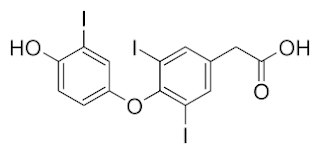
Dilated Cardiomyopathy after Ingestion of TRIAC (triiodothyroacetic acid, Tiratricol)
Donald H. Marks, M.D., Ph.D.


Cardiomyopathy associated with hyperthyroidism is a serious and potentially lethal condition. We describe a patient who developed a dilated cardiomyopathy (DCM) after consuming an over-the-counter (OTC) supplement containing triiodothyroacetic acid. With early diagnosis and treatment by removal of the source, cardiomyopathy may be reversible, as in this patient's case. The dangers of use of OTC medications, and problems and assumptions associated with FDA monitoring and removal from the market of harmful substances are discussed.
Introduction
Tricana, containing triiodothyroacetic acid, was sold as an OTC herbal supplement preparation, and promoted for potential beneficial metabolic effects, including weight loss and energy enhancement. Tricana was withdrawn from the market in 2000 by the FDA 1
when Tricana use was associated with various adverse effects, including heart attacks and strokes. Symptoms associated with ingestion of Triac included many of those typical of hyperthyroidism: insomnia, nervousness, sweating, and diarrhea This report describes the case of a reversible dilated cardiomyopathy in a woman who used Tricana.

Citation:
Donald H. Marks: Dangers of OTC Herbal Supplements: Dilated Cardiomyopathy after Ingestion of TRIAC (triiodothyroacetic acid, Tiratricol). The Internet Journal of Endocrinology. 2007. Volume 3 Number 2.

Friday, October 19, 2018

Interferon and Risk for Drug-Seeking Behavior

Interferon and Risk for Drug-Seeking Behavior

Donald H. Marks M.D., Ph.D. Department of Medicine, Cooper Green Mercy Hospital
Jesse Milby Ph.D. Department of Psychology, University of Alabama at Birmingham

The Internet Journal of Pain, Symptom Control and Palliative Care ISSN: 1528-8277

Citation: D.H. Marks & J. Milby: Interferon and Risk for Drug-Seeking Behavior. The Internet Journal of Pain, Symptom Control and Palliative Care. 2009 Volume 6 Number 2 Keywords: drug craving, drug seeking behavior, interferon, medication, methadone, cocaine

Abstract
Interferon (IFN), a biological medication used to treat viral hepatitis and certain cancers, has clinically significant potential to cause a wide range of adverse neuropsychiatic effects. The spectrum ranges from agitation, aggression, insomnia and irritability, to suicidal thought and drug-seeking behavior (DSB). Out of a total population of 353 patients infected with hepatitis C virus (HCV), 132 patients at an inner city hepatitis clinic underwent treatment. Those treated were questioned at intake and on a regular basis for the initiation or increase of DSB. In addition, when warranted, patients were tested for the presence of drugs they were not prescribed. Over a four year period, ten patients (4 currently receiving treatment with IFN, 4 with prior treatment, and 2 who never received IFN) reported an increase in DSB. The danger of developing IFN-induced DSB appears to be relatively low (< 3%) in our patient population, and we also observed DSB in HCV patients who were not treated with IFN. To our knowledge, this is the first study of the incidence of DSB associated with IFN treatment for HCV that completely surveyed the HCV-treated population and used urine toxicology to verify illicit drug use.

Introduction
Patients with DSB have an inappropriate focus on obtaining a desired abused drug, i.e. cocaine, opioids, methamphetamine, etc. without concern of other more appropriate issues, such as diagnosis or treatment of their addictive behavior (Vissers, 2002). DSB includes abused drug preoccupation (talk and memories), and the craving and actual search for and use of abused drugs. It may be true that DSB as an observed action may not always be preceded by drug-related thoughts and memories as associated cognitive activity. However, it is likely that in most cases these cognitive activities precede DSB as action. Since DSB and use has the potential for dire consequences to IFN therapy in the treatment of HCV, we have questioned for the self-report of such drug abuse-related cognitive activity in regular office visits. As a general rule, it is reasonable to urge patients to avoid any stimulus to DSB while undergoing treatment for viral hepatitis, because of the potentially harmful effects of non-prescribed and addictive medication on antiviral therapy. Since the principle routes of contraction of infection for HCV include injection drug use (IDU), any increase in DSB caused by a side effect of a therapeutic (such as IFN) could become counter- therapeutic. IDU is known to suppress the immune system in some [for example, HIV] viral infections (Thompson & Salvato, 1998). Even though frequent illicit drug use during treatment of HCV may lead to decreased adherence (Sylvestre & Clements, 2007 ), several researchers (Robaeys & Buntinx, 2005); (Sylvestre, Litwin, Clements, & Gourevitch, 2005); (Sylvestre & Clements, 2007 ); (Grebely et al., 2007 ) have reported that illicit drug use itself may not counter the therapeutic response to IFN. Although the Warnings Section, Neuropsychiatric Subsection of the prescribing information (PI) for pegylated interferon alpha 2 (IFN) mentions that “relapse of drug addiction” may occur in patients, no specific information is given on the frequency of occurrence, the causal relatedness or predisposing or inciting factors. Upon query to the manufacturers, they were not able to supply specific data in these areas. A search of Medline also did not reveal specific published information in this regard. The aim of this observational study was to determine the incidence of DSB in a population being treated for HCV in a inner city community hospital, using standardized and previously validated approaches.

Accutane: Focus on Psychiatric Toxicity and Suicide

Accutane: Focus on Psychiatric Toxicity and Suicide
Chapter 20
Donald H. Marks, M.D., Ph.D. and Tzarina Middlekoop, Ph.D.


20.1 Introduction Since early this century, animal research revealed modifications of epithelial structure, such as increased epidermal keratinization and squamous metaplasia of the mucous membrane, under conditions of vitamin A deficiency. The finding that these defects could be corrected by administering vitamin A lead to the emergence of vitamin A as an anti- keratinizing factor. The first synthesis of vitamin A fifty years ago opened a new era into the chemical synthesis of vitamin A derivatives, collectively known as retinoids. First synthesized in 1955, Accutane (Ro 4-3780, isotretinoin), a first generation retinoid, was shown to be highly efficacious in the therapy of disorders of keratinization (e.g., Dariers disease, ichthyosis). Peck was the first investigator to demonstrate this drug’s value in the treatment of severe acne and in September of 1982, it was approved for use in the U.S. by the Food and Drug Administration (FDA). Accutane is Roche’s second biggest seller, generating about $300 million in sales through the first half of 1998. From 1993 to 1997, prescriptions in the U.S. jumped 52 percent (to 1.5 million). 
20.2 Mechanism of Action Acne is due to an interaction of the normal skin bacteria with the patient’s abnormal type of sebaceous lipids, and is associated with an increased sebum production and ductal cornification. The acne bacteria, Propionibacterium acnes reside on the surface of the skin in quite high numbers, especially in oil-rich areas. If they colonize the pilosebaceous duct in the presence of comedones (blackheads and whiteheads), t hen inflammation is likely to be triggered, resulting in papules, pustules and, if inflammation is more expansive, nodules. Although the exact mechanism of the anti-acne action of isotretinoin is unknown, it is unique in its ability to a ffect—albeit not to the same degree—all the known etiological factors of acne, including reduction of sebum production, lessening of comedogenesis, and the decrease of surface and ductal colonization by Propionibacterium acnes. 3

Depression Leading to Suicide As An Adverse Effect of Metoclopramide

Depression Leading to Suicide As An 
Adverse Effect of Metoclopramide

The Internet Journal of Gastroenterology 2007 : Volume 5 Number 2

by Donald H. Marks M.D., Ph.D.

Citation: D. H. Marks : Depression Leading to Suicide As An Adverse Effect of Metoclopramide . The Internet Journal of Gastroenterology. 2007 Volume 5 Number 2
http://ispub.com/IJGE/5/2/10427

Abstract
Metoclopramide (Reglan) is a substituted benzamide derivative with therapeutic utility as a stimulant of upper gastrointestinal motility. Metoclopramide is prescribed widely for diabetic gastric stasis and nausea. Although many of metoclopramide's adverse effects are appreciated by both prescribing physicians and patients, depression can occur, sometimes with devastating results. We describe a case of suicide which was causally related to metoclopramide.

Report of a Case
The patient was a 70 year old Caucasian man who took metoclopramide for the nine months prior to his death from suicide. He had a two year history of documented sliding hiatal hernia with moderate gastroesophageal reflux; an upper GI study showed transient hold up of the barium pill at the level of a hiatal hernia. Medical history included hypertension, hyperlipidemia, bronchiectasis, chronic dysphagia, GERD, hiatal hernia, sick sinus syndrome/atrial fibrillation, and benign prostate hypertrophy. Thyroid panel was normal. The patient's father committed suicide when the patient was 4 years old, and there is little information about this, although the patient's widow thought that there were severe financial pressures involved. The patient did not smoke or use alcohol.

Metoclopramide was prescribed for difficulty swallowing food. Concurrent medications at that point were Zocor, Prenivil, and Protonix. The patient first complained to friends of depression on the third month of treatment with metoclopramide. At the sixth month of metoclopramide treatment, an antidepressant (Zoloft) was prescribed. There was no improvement in his depression. Following the use of Zoloft, three other antidepressant drugs (Prozac, Effexor and Celexa) were tried, also without improvement in his depression. The patient's depression became severe, he lacked motivation to do any physical activity, was increasingly tired, did not desire to interact with others, and slept excessively. According to his wife, the patient had been energetic and optimistic for the 47 years of their marriage prior to the start of metoclopramide. The patient's treating physician stated that his differential diagnoses included Parkinson's vs. Parkinson's with atypical depression pattern vs. early dementia. Thyroid levels were normal. The patient did not exhibit symptoms of akathesia, agitation or flat affect. No cognitive deficits were measured or noted.

Upon referral to a neurologist, it was determined that these symptoms were most likely secondary to metoclopramide use, that the patient was significantly depressed and needed psychiatric follow-up. No mention was given of the danger of suicide or need for protection. A few days later, the patient committed suicide by gunshot to the chest.

Methods
The basis of this case report was a thorough review of the medical records, an interview with the patient's spouse, and a search of the current medical literature using Medline.

Results
Using universally accepted algorithms for the determination of causal relatedness between medication and adverse effects, 1 metoclopramide was determined to be causally related to this suicide. The key bases for this association were: Temporality: Lack of pre-metoclopramide depression or suicidal thoughts, Temporality: Depression and suicide occurred after the start of metoclopramide, Known adverse effect: depression (prescribing info), suicide ideation (peer-reviewed medical literature), Biological Plausibility: Metoclopramide has centrally nervous system actions, and is an antagonist of dopamine, Biological ispub.com/.../depression_leading_to_s... 3/9 5/26/2009 nervous system actions, ISPUB and - Depression is an antagonist Leading to of Suicide... dopamine, Biological Coherence: Does not conflict with what is known, The absence of an alternative explanation.

Discussion
The wide variety of both desired and adverse effects from metoclopramide stem from its ability to act both centrally (nausea) and peripherally (gastric motility), as an antagonist of dopamine, and sensitize gastric smooth muscle to the effects of acetylcholine stimulation. The CNS side effect profile of metoclopramide is broad, and includes drowsiness, extrapyramidal syndrome (dystonias, akathesia), depression, dizziness and insomnia. 2 , 3 , 4

Metoclopramide is similar to two other benzamides - sulpiride and amisulpiride, which are antipsychotics available in England. In fact, metoclopramide itself has clinical antipsychotic efficacy. 5 Antipsychotic treatment has been identified as one of the factors responsible for the increased rate of suicide in schizophrenics, 6 so it follows that any drug with antipsychotic efficacy, and which can cause akathesia (such as metoclopramide) may cause an increased risk of suicide. The current prescribing information for metoclopramide includes a WARNING which states, “Mental depression has occurred in patients with and without prior history of depression. Symptoms have ranged from mild to severe and have included suicidal ideation and suicide. Metoclopramide should be given to patients with a prior history of depression only if the expected benefits outweigh the potential risks.” 2

Clinicians have reported that akathesia can exacerbate psychopathology. 7 It is recognized that akathesia can be linked to both suicide and violence. 9 A link between akathesia and violence, including homicide, following antipsychotic use has also previously been reported. ispub.com/.../depression_leading_to_s... 4/9 8 , 10 , 11 , 12 5/26/2009 ISPUB - Depression Leading to Suicide...

Weddington and Banner identified a patient who developed an organic affective syndrome after administration of metoclopramide. 13 The syndrome was characterized by dysphoria, akathesia, depressed mood with suicidal ideation, insomnia, racing thoughts and labile affect. The symptoms increased within 30 minutes of each dose, and upon discontinuation of metoclopramide, the symptoms gradually resolved (challenge – dechallenge response). Although there have been perhaps eight case reports in the medical literature of depression thought to be causally related to metoclopramide use, to my knowledge this is the first published case of completed suicide caused by metoclopramide.

The DSM-IV defines a Substance-Induced Mood Disorder (SIMD) as having evidence from the history, physical examination or laboratory findings of symptoms developing during, or within a month of substance (in this case, metoclopramide) intoxication or withdrawal, or the medication use is etiologically related, does not occur exclusively during the course of a delirium, the symptoms cause clinically significant distress or impairment in social, occupational, or other important areas of functioning, with Depressive Features: if the predominant mood is depressed. Manic mixed features can also occur. 8

Clearly, from this definition, the patient reported here had experienced a SIMD from metoclopramide toxicity. The patient did not experience a beneficial antidepressive response after treatments with three different antidepressants of the selective serotonin reuptake inhibitor (SSRI) class. This is not completely surprising, in that clinical experience and a number of recent articles point to a disappointing overall beneficial clinical response rate to SSRIs, only slightly greater (57.5%) than for placebo. 14 Increasing the dosage of ispub.com/.../depression_leading_to_s... SSRIs as a means to increase the clinical response often only leads to 5/9 5/26/2009 ISPUB - Depression Leading to Suicide... SSRIs as a means to increase the clinical response often only leads to an increase in adverse effects, and (frequent) switching between SSRIs can lead to its own group of problems (discontinuation syndrome, toxicity).

Fisher and David reported that metoclopramide can interact with SSRI drugs to cause a Serotonin Syndrome. 15 This syndrome results from excessive stimulation of central and peripheral serotonergic receptors, and is characterized by changes in mental status, and motor and autonomic function. SSRI drugs are commonly used antidepressants, which have also been associated with agitation, aggression, and suicide. 2 The two patients reported by Fisher and David were stable on SSRIs before the addition of metoclopramide, 15 whereas in the case reported here, SSRIs were given after depression had arisen from use of metoclopramide. There is no indication that the subject of the current report had developed a Serotonin Syndrome. SSRI medication has clearly been linked with suicidal ideation and suicide. 16 , 17

During the first month of therapy, SSRI antidepressants in patients 66 years of age and older were associated with a nearly fivefold higher risk of completed suicide than other antidepressants. 18 It is possible in this reported case that the addition of SSRI medications may actually have exacerbated the metoclopramide-induced depression and precipitated the suicide. This is why prescribing physicians must be aware of the adverse effect potentials for all medications prescribed for patients, including drug-drug interactions; and must adequately warn their patients and patient families to anticipate problems and what actions to take. In this reported case, depression was caused by metoclopramide, and the appropriate treatment would have been discontinuation of .....

MR imaging of Drug-induced Suicidal Ideation

MR Imaging of Drug-Induced Suicidal Ideation

D Marks, M Adineh, S Gupta
 The Internet Journal of Radiology. 2007 Volume 9 Number 1.
Abstract
Two patients with a history of suicidal ideation (SI) underwent functional MR imaging while undergoing treatment with interferon alpha 2 (IFN) for Hepatitis C virus infection (HCV). Patient #77 had a remote history of SI, but no current SI when treated with IFN. Patient #288 experienced an IFN-heightened SI, although she denied intent, a plan or a means. Visual stimuli were presented during functional MR imaging (fMRI) that were designed to invoke thoughts of suicide and violence. Patient #77 showed activation expected for visual stimulation alone, whereas patient #288 showed heightened activation for some of the visual stimuli with violent emotional content. Functional MR imaging shows promise to screen for a number of medication-induced CNS adverse effects (AE).
 

Introduction

Treatment of infections such as HCV with currently available medications can be associated with serious medical/clinical consequences, including psychological sequellae (prescribing information - PI). The use of interferon has been associated with SI (PI; Janssen et al., 1994; Fukunishi et al., 1998; Schafer et al., 2000; Ademmer et al, 2001; Bagheri et al., 2004; Dieperink et al., 2004; Laguno et al., 2004), although the incidence is not well-characterized (Helbling et al., 2002). The rate at which drug-induced SI progresses to a suicidal act is also not known. Other than avoiding use of IFN in individuals with significant underlying depression (Barraclough et al., 1974) or other risk factors for suicidal thought (CDC 2007), there is currently no reliable way to prevent an individual from responding to IFN by developing SI. It will therefore be of immense value to be able to predict and monitor in a quantifiable manner a clinically significant suicidal response to IFN in patients prior to the initiation of treatment, and also during the drug development cycle.

Functional neuroimaging has been successfully applied to the study of mood-disorders, including endogenous [Fu et al et al., 2004] and medication-induced (Marks et al., 2007a) depression, anxiety, and drug-seeking behavior. The application of fMRI for SI follows logically (Mann 2005). We have previously demonstrated (Marks et al., 2007a ) interesting differences between MR imaging of major depressive disorder (MDD) and that of medication-induced depression and anxiety, implying that the underlying causative mechanisms and treatment may differ. This report describes an initial effort to adapt fMRI to identify and monitor the course of SI induced by medications. This approach holds the potential of introducing new paradigms for understanding and treating SI.

Methods

Monday, October 15, 2018

Face Recognition, Reversible Correlation Between fMRI and Biometrics Data

Face Recognition, Reversible Correlation Between fMRI and Biometrics Data
D H Marks, A Yildiz, S Vural, S Levy
Citation:  D H Marks, A Yildiz, S Vural, S Levy. Face Recognition, Reversible Correlation Between fMRI and Biometrics Data. The Internet Journal of Radiology. 2017 Volume 20 Number 1.
Abstract
Specific individual face recognition in the brain has been demonstrated with analysis of three dimensional neural activation patterns – cognitive engrams – revealed by functional magnetic resonance imaging (fMRI). Individual faces can also be differentiated by biometric pattern recognition from camera images using biometric analysis. A correlation between face recognition data obtained from these two methods is now documented. A two way correspondence between face data obtained by these and other means exists, which should facilitate face recognition, the utility of interrogation, and further the understanding of cognition.
INTRODUCTION
Since 2006, it has been known that widely arranged brain cortical response patterns elicited by individual face images with high-resolution functional magnetic resonance imaging (fMRI) can be used to discriminate between unique faces (1). This work has been independently validated by other research laboratories (2,3). Face activation patterns obtained by fMRI are known to be related to and vary with the structure of the face (4) and these variations are consistent across individuals. Cognitive engrams refer to multi-dimensional representations of brain activation in response to specific stimuli (1). Cognitive engrams can be arranged into a [Rosetta] database which relates the Cognitive Engrams and other associated data to specific mental concepts, i.e., a visual representation of actual memory patterns. Faces can also be analyzed and correlated with their physical features (5,6,7). Relative sizes and distances for facial landmarks such as the eyes, nose, ears, chin, and skin texture, among others can be measured. Face data can be extracted from camera images, or from video streams. Principle methods for biometric face analysis are geometric, which is feature based, and photometric, which is view based. Many different algorithms for face analysis have been developed, including principal component analysis PCA, linear discriminant analysis LDA, elastic Bunch graph matching EBGM, and more recently deep-learning (DL) based non-linear feature extraction methods. Face structure, as are all of our physical characteristics, are coded within DNA. Claes et al. (8) used extensive modeling methods to determine the relationships between facial variation and the effects of sex, genomic ancestry, and a subset of craniofacial candidate genes. Their modeling could lead to approximating the appearance of a face from genetic markers alone. Knowing that face recognition by fMRI and by biometrics both depend on the physical differences between individual faces, a correlation of face recognition from these two different data sources was studied and reported herein.
METHODS
Overall, the steps used in this study are: Test subjects view pictures of face, object, or concept, or has other visual or auditory stimulation, while undergoing functional neuroimaging, Functional neuroimaging data is collected, 3-D Activation map is constructed, which constitutes the specific Cognitive Engram for the face / object imaged, Collection of activation maps is added to a (Rosetta) Database of activation maps The same faces used for generating fMRI activation maps are examined by (video) camera, and a biometric analysis is generated, One-to-one correspondence is made between the fMRI activation map (the facial cognitive engram) and the biometric data.
PREPARATION OF FUNCTIONAL MR IMAGING ACTIVATION MAPS / COGNITIVE ENGRAMS OF FACES
3-D activation maps using fMRI can be prepared, as previously described (1). Briefly, normal volunteers are shown faces by rear projection screen or other methods (such as video projection goggles) while the test subjects are undergoing functional MR imaging. To perform fMRI, each test subject lays within a GE Cigna 3-T Signa 11X Excite MRI scanner, wearing a phased array whole head coil, mounted with a 45 degree mirror. This arrangement allowed test subjects to see images displayed onto a rear projection screen positioned by their feet. fMRI was performed while viewing the test stimuli it order to capture functional data, as described by Marks et al (1). A short localizer MRI scan was performed to verify that the field of view was within the skull, and to assure the absence of “ghost” images. A high-resolution full volume structural MRI scan was then obtained for each subject, using fast SPGR imaging (146, 1.0-mm thick axial slices, no spaces, TR = 8, TE = 3.2, FOV = 24 cm, 256 £ 256 matrix). These T1-weighted images provided detailed anatomical information for registration and 3-D normalization to a standard atlas. Test subjects were then shown photos / images generated by PC using PowerPoint (Microsoft) and projected onto a rear projection screen placed at the foot of the test subject, as described in Marks et al (1). Pictures were viewed by means of a mirror system mounted on the head coil. Changes in the blood oxygen level dependent (BOLD) MRI signal were measured using a gradient-echo echoplanar sequence. The following sequences were used, but variations are available. Continuous fMRI scans lasted 110 seconds each. EPI parameters were: TE 35, TR 2000, multiphase screen, 55 phases per location, interleaved, flip angle 90, delay after acquisition-minimum. Using a visual stimulus package, color photographs were presented in a mini-block design while neuroimaging was performed. In a typical session, after a 4 second lead-in time, a blank screen was displayed for 4 seconds, then the picture of interest for 4 seconds, and this was repeated for the scan time. The fMRI scan volumes were motion-corrected and spatially smoothed in-plane. MRI data files were normalized and analyzed using MedX (version 3.4.3, Sensor Systems, Sterling, VA) to compute statistical contrasts and create a map representing significantly activated areas of the brain that responded differentially to a visual test stimuli. For the voxels that show an overall increase in activity for meaningful stimuli, a positive regression analysis for the contrast between a test photo and control (blank page) stimuli was conducted, creating an activation map containing specific voxels with an uncorrected probability, P ≤ 0.05; meaning every voxel showing activation with the probability greater than 95%. Only those activated voxels were selected for further analysis. That statistical map was then superimposed on coplanar high-resolution structural images. The partial volume structural images were registered with the full volume high-resolution images using Automated Image Registration (9). Those full volume high-resolution images were then transformed (registered and normalized) to the Talairach and Tournoux atlas (10) using MedX tools. Each activated voxel on these images was selected to obtain Talairach coordinates of brain regions that respond maximally to the test stimuli and to further generate a Cognitive Engram. Comparison of observed patterns of activation were correlated with the nature of the response, such as face recognition, or a truthful or deceptive response.
Three dimensional graphical representations of the identified activation maps were constructed by plotting the xyz coordinates, using the program DPlot (HydeSoft Computing, Vicksburg, MS).
PREPARATION OF BIOMETRIC DATASETS OF FACES
The same photographs of faces used to prepare fMRI data were then introduced to a biometric system. Ayonix Corporation (Tokyo, Japan) software was used, but other commonly available biometric systems should work as well. Biometric face data sets were then generated. The Ayonix face extraction model uses customized HOG- like (Histogram of Oriented Gradients) features on a high number of overlapping face regions to reduce the effects of noise, viewing angle, aging, facial expressions and occlusions. This software extracts features from the whole face together, however, each feature window is aligned onto one of the facial landmark locations extracted in the pre- processing step. This way, the Ayonix software encodes both local and global information about the face geometry and appearance. The Ayonix software is constructed by training models on hundreds of thousands of faces from different age groups, genders and races, with different viewing and lighting conditions; thus appearance differences between Face Recognition, Reversible Correlation Between fMRI and Biometrics Data 3 of 7 different groups of people (ie. European and Asian) are Figure 1 inherently encoded in the feature extraction step of that software. Skin color is not taken into account with the Ayonix software, and the images are converted to grayscale before processing. Following is a general outline of steps used to create face biometric data sets using Ayonix software: Step 1. Face pre-processing - Face is detected by face detection engine - Face region is cropped and resized to a fixed image - Face region is converted to grayscale - Facial landmarks are extracted on the face - Geometric alignment is performed - Lighting effects are corrected - Facial quality is measured Step 2. Face feature extraction - Face features are extracted from overlapped areas on detected facial landmarks - Feature normalization and corrections are performed - Features are transformed into recognition space using the Ayonix engine
CREATION OF CORRESPONDENCE GRID:
A one-to-one correspondence grid was constructed (Figure 1 and Table 1). The two components were fMRI consensus activation points and biometric face data, arranged by each of the five static faces used in this study. Correspondence was made using regression analysis and other mathematical analyses.
RESULTS Using data from fMRI and biometrics, a one to one correspondence grid was constructed. A graphical illustration of the process is shown in Figure 1. The correspondence grid is shown in Table 1. The data for fMRI was previously provided (1). The face biometric data sets are embedded into Figure 1. Table 1 is a table of data in various formats illustrating how correlated data on brain activation obtained from functional MR imaging while viewing specific visual stimuli correlates with the biometric data obtained using facial recognition software. Table 1 Table 1 is a table of data in various formats illustrating how correlated data on brain activation obtained from functional MR imaging while viewing specific visual stimuli correlates with the biometric data obtained using facial recognition software.
DISCUSSION A wide multivoxel activation pattern (1,17) is seen with fMRI during object and face recognition. As noted in prior publications, and in an issued patent (18), these object- specific or concept-specific activations are referred to as Cognitive Engrams. Cognitive Engrams may reflect neuronal population codes (1,17,18). Marks et as and Muir Face Recognition, Reversible Correlation Between fMRI and Biometrics Data (17) have previously shown (1,18) that Cognitive Engrams possess representational content. Various researchers, including Marks (1) and later by Kriegeskorte (2) and others have shown that fMRI activation data can be interpreted to identify individual, specific representational content, such as faces, unique objects, emotions, truthful and deceptive statements to questions, and other cognitive content It is know that face recognition by biometric analysis (5) of a picture of a face is very dependent on the structure of the face. Similarly, there is evidence that face representation by fMRI is also dependent on facial geometry. Loffler et al. (11) found evidence that neural activation patterns for individual faces are encoded as grouped data. This encoding varied on the direction (facial identity) and distance (distinctiveness) from standard or prototypical (mean) face. Loffler et al found that varying facial geometry (head shape, hair line, internal feature size and placement) caused the corresponding fMRI signal to increase with increasing distance from the mean face. Loffler also determined that the same neural population will respond to faces falling along single identity axes within this space. Boccia (12) found that the pattern of activity in most of these areas specifically codes for the spatial arrangement of the parts of the mental image Rotshtein et al (13) found that fMRI of varying facial content demonstrated differential activity in critical face recognition areas of the brain. The inferior occipital gyrus (IOG) showed sensitivity to physical rather than to identity changes. The right fusiform gyrus (FFG) showed sensitivity to identity rather than to physical changes. Bilateral anterior temporal regions show sensitivity to identity change that varies with the subjects' pre-experimental familiarity with the faces. These findings supported differential activity within the brain taking part in distinguishing varied facial content. Rotshtein et al (4) used fMRI to study how the brain processes featural information and second order spatial relations in face identity processing. Features included eyes, mouth, and nose. second-order spatial relations were measured between face features. They found that feature- dependent effects occurred within the lateral occipital and right fusiform regions of the brain. Spatial relation effects occurred in the bilateral inferior occipital gyrus and right fusiform. Overall, Rotshtein et al found that featural and second-order spatial relation aspects of faces make distinct contributions to behavioral discrimination and recognition. 4 of 7 Face features contributed most to face discrimination, whereas second-order spatial relational aspects correlated best with recognition skills. These results support ongoing findings employing fMRI for face recognition tasks. Rotshtein et al (14) then used "hybrid" faces containing superimposed low and high spatial frequency (SF) information from different identities. They found that repetition and attention affected partly overlapping occipitotemporal regions but did not interact. Changes of high SF faces increased responses of the right inferior occipital gyrus (IOG) and left inferior temporal gyrus (ITG), with the latter response being also modulated additively by attention. In contrast, the bilateral middle occipital gyrus (MOG) responded to repetition and attention manipulations of low SF. A common effect of high and low SF repetition was observed in the right fusiform gyrus (FFG). Follow-up connectivity analyses suggested direct influence of the MOG (low SF), IOG, and ITG (high SF) on the FFG responses. Overall, their results showed that different regions within occipitotemporal cortex extract distinct visual cues at different SF ranges in faces and that the outputs from these separate processes project forward to the right FFG, where the different visual cues may converge. These results support ongoing findings employing fMRI for face recognition tasks and illustrate how analysis of differential brain activation demonstrates differential recognition of faces Cohen et al (22) demonstrated that individual face images could be accurately reconstructed from distributed patterns of neural activity, even when excluding activity within occipital cortex. Miyakawi et al (23) and Schoenmakers et al (24) showed that image reconstruction can occur based upon the brain activation pattern data alone, without the need for prior internal pattern references. Nishimoto et al (25) were able to interpret dynamic brain activity (viewing of movies) using a motion-energy encoding model and a Bayesian decoder. In essence, three dimensional activation patterns from wide areas of the brain are formed into patterns which are equated back to the stimulus for the activation (face, object, concept, emotion). Ultimately, all activation patterns in three dimensional space form unique data sets specific for the object or concept under consideration – Cognitive Engrams (1). Analysis of faces by means of biometrics has its own complex art and science, as described elsewhere. Just as with Face Recognition, Reversible Correlation Between fMRI and Biometrics Data visual recognition by the human eye, biometric systems depend heavily on differences in face structure, tone and other characteristics. There are a number of methods to analyze complex fMRI face activation data, including: sparse logistic regression (15), feature vectors (16) using a support vector machine algorithm; quantitative receptive- field models (3), multivoxel pattern information analysis (17), direction and distance from a prototypical (mean) face (11), partial least squares (19), and others. These methods are described in the referenced articles. EVIDENCE FOR REVERSE LOOKUP = MIND- READING, A FORM OF REVERSE LOOKUP = MIND-READING. Cox (20) used multivariate statistical pattern recognition methods, including linear discriminant analysis and support vector machines, to interpret activation patterns of fMRI. Test subjects looked at categories of objects, rather than specific variations within a class. They were able to determine within some degree of experimental error which category (as opposed to unique individual) of object or picture their test subjects were looking at. Thirion (21) used retinotopy of the visual cortex to infer the visual content of real or imaginary scenes from the brain activation patterns that they elicit. Yamashita (15) used a novel linear classification algorithm, called sparse logistic regression (SLR), to automatically select relevant voxels while estimating their weight parameters for classification. Using simulation data, they confirmed that SLR can automatically remove irrelevant voxels and thereby attain higher classification performance than other methods in the presence of many irrelevant voxels. These patterns of activatated voxels formed what can best be described as cognitive engrams, which can be used to predict or decode fMRI activity patterns. SLR also proved effective with real fMRI data obtained from two visual experiments, successfully identifying voxels in corresponding locations of visual cortex. SLR-selected voxels often led to better performance than those selected based on univariate statistics, by exploiting correlated noise among voxels to allow for better pattern separation. Extensive research in the published scientific literature, patent sources and internet indicate that a two-way lookup between fMRI and biometric data is a unique, original and not previously explored approach to face recognition. 5 of 7 This concept further allows the practical interpretation of thoughts, emotions, feelings, intents using neuroimaging data. The reverse lookup concept allows the categorization and compiling of thoughts in the Rosetta Database, and ways to store and retrieve cognitive engrams. Correlation of individual biometrics to specific thought patterns via functional neuroimaging will further the identification of individuals and the interpretation of their associated concepts and intents.
References
1. Marks DH, Adineh M, Wang B, Gupta S, Udupa JK. Multidimensional Representation of Concepts as Cognitive Engrams in the Human Brain. The Internet Journal of Neurology [peer-reviewed serial on the Internet]. 2007. Volume 6, Number 1. 2. Kriegeskorte N, Formisano E, Sorger B, Goebel R. Individual faces elicit distinct response patterns in human anterior temporal cortex. Proc Natl Acad Sci USA. 2007 Dec 18;104(51):20600-5. 3. Kay KN, Naselaris T, Prenger RJ, Gallant JL Identifying natural images from human brain activity. Nature. 2008 Mar 20;452(7185):352-5. 4. Rotshtein P, Geng JJ, Driver J, Dolan RJ. Role of Features and Second-order Spatial Relations in Face Discrimination, Face Recognition, and Individual Face Skills: Behavioral and Functional Magnetic Resonance Imaging Data. J Cogn Neurosci. 2007 Sep;19(9):1435-52. 5. Hammoud RI, Abidi BR, Abidi MA. Face Biometrics For Personal Identification: Multi-Sensory Multi-Modal Systems. Springer, 2007. 6. Turk, Matthew A and Pentland, Alex P. Face recognition using eigenfaces. Computer Vision and Pattern Recognition, 1991. Proceedings {CVPR'91.}, {IEEE} Computer Society Conference on 1991 7. Wing N et al. Biometrics History http://www.biometrics.gov/Documents/BioHistory.pdf 8. Claes P, Liberton DK, Daniels K, Rosana KM, Quillen EE, et al. (2014) Modeling 3D Facial Shape from DNA. PLoS Genet 10(3): e1004224. 9. Woods RP, Mazziotta JC, Cherry SR. MRI-PET registration with automated algorithm. Journal of Computer Assisted Tomography 1993;17:536-546. 10. Talairach, J., & Tournoux, P. (1988). Co-planar stereotaxis atlas of the human brain (M. Rayport, Trans.). Face Recognition, Reversible Correlation Between fMRI and Biometrics Data New York: Thieme Medical. 11. Loffler G, Yourganov G, Wilkinson F, Wilson HR. fMRI evidence for the neural representation of faces. Nat Neurosci. 2005 Oct;8(10):1386-90. 12. Boccia M Piccardi L, Palermo L et al. A Penny for Your Thoughts! Patterns of fMRI Activity Reveal the Content and the Spatial Topography of Visual Mental Images. Human Brain Mapping 36:945–958 (2015) 13. Rotshtein P, Henson RN, Treves A, Driver J, Dolan RJ. Morphing Marilyn into Maggie dissociates physical and identity face representations in the brain. Nat Neurosci. 2005 Jan;8(1):107-13. 14. Rotshtein P, Vuilleumier P, Winston J, Driver J, Dolan R. Distinct and Convergent Visual Processing of High and Low Spatial Frequency Information in Faces. Cereb Cortex. 2007 Nov; 17(11): 2713–2724. 15. Yamashita O, Sato MA, Yoshioka T, Tong F, Kamitani Y. Sparse estimation automatically selects voxels relevant for the decoding of fMRI activity patterns. Neuroimage. 2008 Oct 1;42(4):1414-29. 16. Lee JH, Marzelli M, Jolesz FA, Yoo SS. Automated classification of fMRI data employing trial-based imagery tasks. Medical Image Analysis 13 (2009) 392–404. 17. Mur M, Bandettini PA, Kriegeskorte N. Revealing representational content with pattern-information fMRI--an introductory guide. Soc Cogn Affect Neurosci. 2009 Jan 17. 18. Marks, DH. Brain Function Decoding Process And 6 of 7 System. US 7,627,370, 19. Maurer D, O’Craven KM, Le Grand R, Mondloch CJ et al. Neural correlates of processing facial identity based on features versus their spacing. Neuropsychologia 45 (2007) 1438–1451 20. Cox DD, Savoy RL. Functional magnetic resonance imaging (fMRI) "brain reading": detecting and classifying distributed patterns of fMRI activity in human visual cortex. Neuroimage. 2003 Jun;19(2 Pt 1):261-70. 21. Thirion, B. et al. Inverse retinotopy: inferring the visual content of images from brain activation patterns. Neuroimage 33, 1104–1116 (2006). 22. Cowen, A.S., Chun MM,. Brice A. Kuhl BA, Neural portraits of perception: Reconstructing face images from evoked brain activity. NeuroImage Volume 94, 1 July 2014, Pages 12–22. 23. Miyawaki Y, Uchida H, Yamashita O et al. Visual Image Reconstruction from Human Brain Activity using a Combination of Multiscale Local Image Decoders. Neuron. 2008 Dec 10;60(5):915-29. 24. Schoenmakers S, Barth M, Heskes T, van Gerven M. Linear reconstruction of perceived images from human brain activity. NeuroImage Volume 83, December 2013, Pages 951–961 25. Nishimoto S , Vu AT, Naselaris T, Benjamini Y, Yu B, Gallant JL. Reconstructing Visual Experiences From Brain Activity Evoked by Natural Movies. Current Biology 21, 1641–1646, October 11, 2011. Face Recognition, Reversible Correlation Between fMRI and Biometrics Data Author Information Donald H. Marks, M.D., Ph.D. Millennium Magnetic Technologies, LLC Hoover, Alabama Alparslan Yildiz Ayonix Corporation, The Face Recognition Company Tokyo, Japan Sadi Vural Ayonix Corporation, The Face Recognition Company Tokyo, Japan Steve Levy, MD Westport, Connecticut USA 7 of 7

Monday, October 1, 2018

Documentation of Acute Neck Pain in a Patient Using Functional MR Imaging

Documentation of Acute Neck Pain in a Patient
Using Functional MR Imaging
http: //www,ispub.corn/journal/the internet journal of pain symptom control and~alliat... I/12/2011
The Internet Journal of Pain, Symptom Control and Palliative Care : Volume 8 Number 1
DH Marks, P Valsasina, MA Rocca, M Eilippi
Abstract
Neuroimaging tvas applied to proiride visual documentation ot'the presence of pain in a patient complaining of
long-term neck pain, Activation in the pain-related brain matrix was increased after induced pain, compared
with a resting baseline condition, and decreased after the pain had subsided somewhat.
Introduction
The Internet Journal of Pain, Symptom Control and Palliative Care 2010 :Volume 8 Number 1
Documentation of Acute Neck Pain in a Patient
Using Functional MR Imaging
DHMarks
PValsasina

My Blog List